
Precision Cancer Therapy
Precision Radiotherapy: Innovations in Particle Therapy and Beam Monitoring
Radiotherapy is a cornerstone of modern cancer treatment, with X-rays and gamma rays being the most widely used modalities. However, recent advances have introduced charged particle therapy — using protons and carbon ions — as a highly precise alternative. These particles, accelerated to high energies, deposit their maximum energy within a sharply defined tissue volume (the Bragg peak), minimizing damage to surrounding healthy tissue. By modulating beam energy and direction, clinicians can target tumors with millimeter precision, sparing critical organs.
The Challenge of Beam Monitoring
A key limitation in particle therapy lies not in the beam itself but in the accuracy of tumor localization during treatment. Current beam monitoring systems at facilities like the Heidelberg Ion-Beam Therapy Center (HIT) rely on gas-filled ionization chambers and multiwire chambers (MWCs) to measure dose, position, and beam spot size. However, MWCs cannot operate in magnetic fields, ruling out their use alongside MRI-guided radiotherapy — a critical limitation, as real-time MRI imaging could dramatically improve targeting precision and reduce healthy tissue exposure.
Introducing the MRI-Compatible Beam Monitor
To overcome this limitation, we are developing a next-generation beam monitor based on High-Voltage Monolithic Active Pixel Sensor (HV-MAPS) technology. This innovation enables:
- MRI compatibility: Integration with MRI systems for simultaneous imaging and irradiation.
- High spatial resolution: Precise real-time tracking of beam position, size, and intensity.
- Radiation hardness and ultra-thin design: Critical for operation in high-dose environments while minimizing beam distortion.
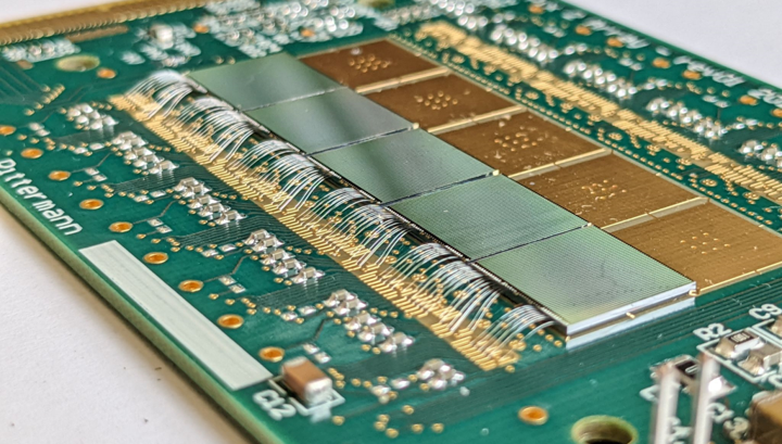
To meet these requirements we have designed HitPix ASIC, a custom-designed sensor chip featuring:
- Pixel size of 200 µm × 200 µm.
- Integrated counting electronics within each pixel.
- Enclosed CMOS transistors for enhanced radiation tolerance.
- High-rate capability (>10 MHz/pixel) to handle intense particle beams.
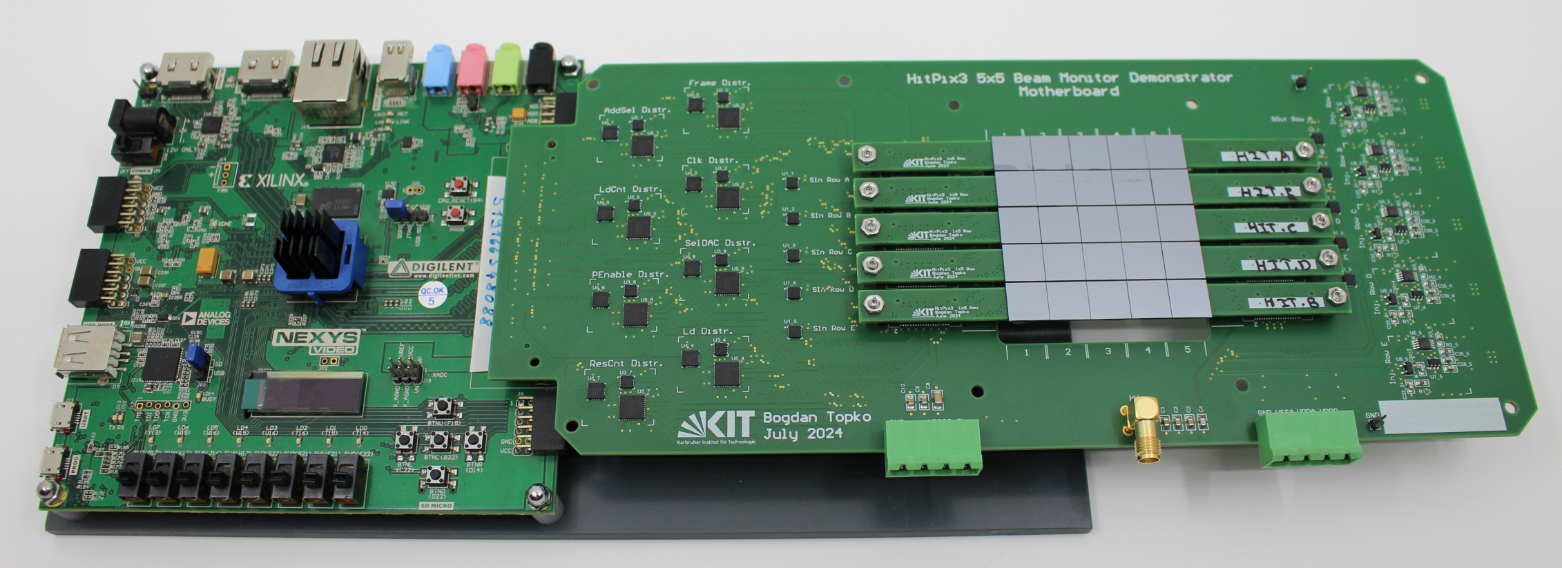
Validation and Collaboration
A multichip detector prototype incorporating HitPix sensors has been successfully assembled and tested in beam trials at HIT. This project, supported by HeiKA (Heidelberg Karlsruhe Strategic Partnership), Helmholtz Programme Matter and Technology and HIT, aims to pave the way for clinical deployment of MRI-guided particle therapy.
Impact and Future Prospects
By enabling real-time MRI during irradiation, HV-MAPS technology could reduce treatment margins, lower radiation exposure to healthy tissue, and improve outcomes for tumors near sensitive organs. The success of HitPix also highlights HV-MAPS as a transformative platform for future detector systems in high-energy physics and medical imaging.